Reversible Chromoplast Transformation: Plastid Interconversion and Greening in Light
 libre
libre
Chromoplasts can convert back into chloroplasts. This reversibility of plastid transformation — long debated since the nineteenth century — has now been demonstrated across many plant tissues using electron microscopy, overturning the older view that chromoplasts were merely degenerating, dead-end organelles.
In 1883 A. Meyer, studying the plastids of carrot roots, first described the greening of chromoplasts in light. A. Schimper established that leucoplasts, chloroplasts and chromoplasts are in fact homologous cell organelles capable of interconverting into one another.
From that point on the idea of the reversibility of plastid metamorphosis — including the ability of chromoplasts to turn into other plastid types — became widely accepted. However, studies carried out with electron microscopy revealed a similarity between the ultrastructural organization of globular-type chromoplasts and the plastids of yellowed leaves.
Why were chromoplasts once thought to be degenerating organelles?
Chromoplasts were once regarded as a terminal, degenerative stage of plastid development because their structure resembled that of plastids in yellowing leaves. Since those leaf plastids form from the chloroplasts of green leaves through degradative processes, A. Frey-Wyssling and his followers concluded that chromoplasts, by analogy with the plastids of yellowed leaves, are a product of the degeneration and breakdown of functional plastids and are therefore incapable of converting into leucoplasts or chloroplasts.
These authors went further, claiming that chloroplasts cannot turn into leucoplasts, and proposed a monotropic (one-directional) transformation of plastids according to the following scheme: 
The view of chromoplasts as plastids in a state of breakdown found support among specialists and was reflected in numerous manuals, reference works and other studies on the ultrastructure of plant cells.
What evidence overturned the one-way model?
Direct observations of intermediate plastid forms refuted the one-way model. W. Straus, studying the plastids of carrot roots, observed transitional forms between chromoplasts and chloroplasts after the roots were illuminated, thereby confirming the conclusions reached by A. Meyer and A. Schimper, while F. Schötz and F. Senser described a reversible transformation of plastids in mutant forms of evening primrose (Oenothera).
The possibility of the conversion of chromoplasts into chloroplasts was subsequently demonstrated in many organisms using electron microscopy.
Plastids of yellow-green fruit
The peel of oranges grown in southern California reaches its maximum orange coloration in the winter months and later (in spring and summer) turns green. The degree of greening is influenced by temperature and nutritional conditions, as well as by potassium gibberellate.
W. Thomson and colleagues studied the ultrastructural changes in plastids during the greening of ripe orange peel after the fruit was immersed in a potassium gibberellate solution. The chromoplasts of the orange fruit contained a large number of electron-dense plastoglobules (800 nm in diameter) with a poorly developed membrane system. 
Small vesicles were also noted, most likely formed from the inner envelope membrane. Another portion of vesicles was visible free in the peripheral regions of the stroma, while some vesicles were in direct contact with grana membranes.
The plastids of light-green fruit had well-developed grana, although individual plastids varied considerably in the degree of development of the internal membrane complex. In some organelles the internal membrane system was poorly developed, while in others it occupied most of the plastid volume.
The plastids of light-green fruit contained a large number of electron-dense plastoglobules. Orange fruit treated with potassium gibberellate acquired a darker coloration by the end of the experiment (four months after treatment), though not as deep as in unripe fruit.
Plastids at this stage showed great structural diversity. Many were identical to the plastids of light-green fruit, with a well-developed membrane system and large electron-dense plastoglobules. Other organelles were structurally indistinguishable from the chloroplasts of deep-green unripe fruit.
In these plastids the number and size of plastoglobules were markedly smaller, and they were characterized by a more developed membrane system. On the basis of their research, W. Thomson and colleagues concluded that during the greening of orange fruit in storage, chromoplasts are converted into chloroplasts.
They note that the appearance of typical chloroplasts in the greened fruit cannot be explained by their formation from proplastids, because in stored fruit with orange, yellow-green, light-green and green coloration no proplastids with a structure characteristic of transitional forms between proplastids and chloroplasts were found. 
The authors also concluded that the membranes of the grana and intergranal elements form through invagination of the inner envelope membrane, regarding the reversibility of chromoplast metamorphosis as a function of the inner envelope membrane.
Taking into account earlier publications on the irreversibility of the plastids of old yellowing tree leaves (in particular the work of S. Toyama and R. Ueda) and classing these organelles as chromoplasts, W. Thomson and colleagues suggested that the final irreversibility of chromoplast metamorphosis occurs when, as a result of changes in the structural and chemical properties of plastid membranes, those membranes lose the ability to form vesicles.
The authors also believe that the formation of the internal membrane system during the establishment of chloroplasts takes place using material from the electron-dense plastoglobules of the chromoplasts.
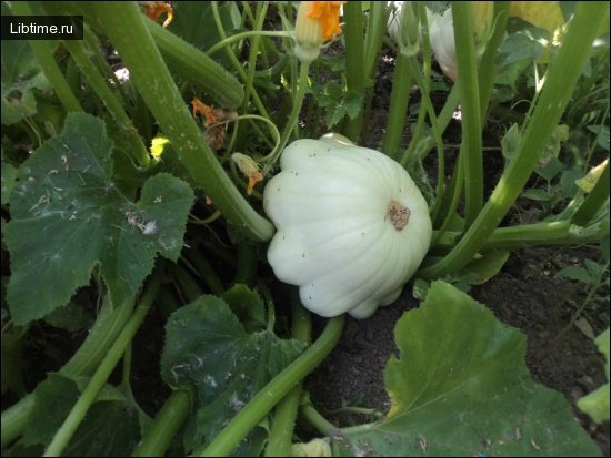
Greening watermelon rind: chromoplasts becoming chloroplasts
B. T. Matienko and co-workers studied changes in plastid ultrastructure in the subepidermal cells of yellow-orange fruit of the table watermelon variety Zheltokoryi. These fruit were characterized by broad yellow-orange stripes on the rind that turned green during storage. In the subepidermal cells of the yellow-orange regions of the fruit, chromoplasts rich in osmiophilic globules (plastoglobules) were found.
There were also small osmiophilic globules with a rounded or oval outline, as well as large osmiophilic formations with a lobed, divided and dissected outline. The plastoglobules were grouped together or diffusely scattered throughout the plastid stroma. Contacts between the osmiophilic globules and the bounding membranes of vesicles were quite frequently observed.
The matrix of large chromoplasts was an electron-transparent mass, while that of small ones had a comparatively higher electron density. In addition to these plastids with a typical chromoplast organization, the authors also found, in the cells of the yellow-orange regions of the fruit, plastids with an intermediate structure (chlorochromoplasts or chromochloroplasts).
In the plastids with an intermediate organization there were vesicles and an increased number of chromolipid globules normally characteristic of chromoplasts, as well as individual grana made up of weakly expressed thylakoids resembling those of chloroplasts, together with an increased electron density of the matrix. When contrasted with uranyl acetate and lead citrate, structures resembling ribosomes were seen, as well as electron-dense lobed formations.
Typical chromoplasts and plastids with an intermediate organization were distributed in the cells of the subepidermal layer in a definite pattern. Most often they occurred in the same cell, regardless of the depth of that cell within the subepidermal layer.
Typical chromoplasts, however, were confined mainly to cells located in the inner part of the subepidermal zone, further from the fruit surface. In the outer cells there were fewer typical chromoplasts and more plastids with an intermediate organization.
The plastids of the subepidermal cells in the greening regions of the rind resembled the chromoplasts and chromochloroplasts of the yellow-orange regions. There were, however, some differences: inside the plastids of the greening regions, the masses of lobed formations split into layers and formed thylakoids similar to those of the grana of green plastids. Groups of thylakoids were also observed in the stroma, arranged as in green plastids.
These organelles had fewer vesicles than chromoplasts. Inside the plastids of greening regions with a more intense green color, the authors observed the complete disappearance of the lobed osmiophilic formations and the formation of 4–5 or more grana in their place. As the green color intensified, the number of thylakoids in the plastids increased and bodies resembling polyribosomes formed.
The polysome-like formations were usually concentrated in particular regions of the chromoplasts that had a more electron-transparent content. Apart from the polysome-bearing areas, individual ribosome-like granules were scattered throughout the chromoplast matrix. In the electron-dense lobed osmiophilic formations, double contrasting increased the electron density, indicating intensified processes of biosynthesis of nucleic acids, proteins and other substances, including green pigments.
The authors believe that the structural changes during the metamorphosis of chromoplasts into chloroplasts are governed by, and accompanied by, a whole series of biochemical rearrangements. In particular, it was shown that the appearance of lamellae and individual grana is accompanied by an increase in the content of chlorophylls a and b in the plastids during greening. The amount of carotenoids also increased at the same time.
Analyzing the results obtained, B. T. Matienko and colleagues concluded that during the greening of the yellow-orange regions of watermelon fruit in storage, chromoplasts are converted into chloroplasts. The authors note that the bulk of the plastids in the yellow-orange regions are represented by chromoplasts rather than intermediate plastids.
At the same time, in the greened regions neither chromoplasts nor the products of their degradation were found. All plastids at this stage contained grana in place of the lobed formations and were represented by typical chloroplasts. Z. Devidé and N. Ljubešić described the conversion of chromoplasts into chloroplasts in the subepidermal tissue of pumpkin.
Conversion in pumpkin tissue
The cells of the dark meridional zones contained typical chloroplasts with a high chlorophyll content and high photosynthetic activity, while the cells of the light zones contained less developed chloroplasts with fewer thylakoids, a low chlorophyll content and lower — but still significant — photosynthetic activity.
At the start of fruit storage, both types of chloroplast develop into typical chromoplasts containing a dense mass of plastoglobules and rare, barely noticeable fragments of thylakoid remnants. These organelles contained no chlorophyll at all and had no photosynthetic activity. After six months of storage the fruit were exposed to light.
As a result, some plastids changed color from orange-yellow to greenish, regardless of whether they were located in a dark or a light zone of the fruit. The chlorophyll content increased and photosynthetic activity was restored. In sections, successive transitional forms from chromoplasts to chloroplasts were observed. In the yellow-orange stripes, however, neither proplastids nor organelles with remnants of chloroplast structures were found.
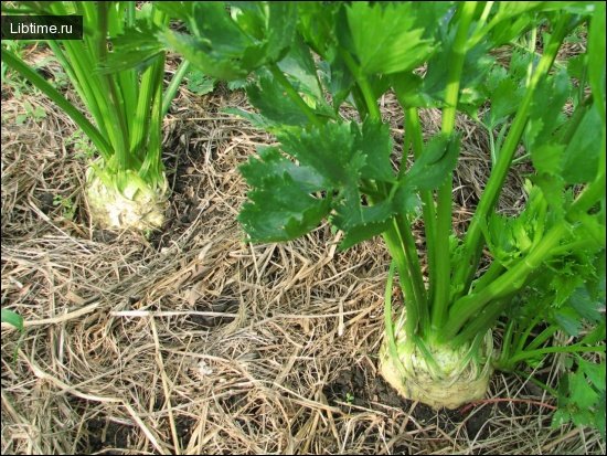
It was established that in the subepidermal cells of pumpkin a transformation of chromoplasts into chloroplasts takes place. P. Grönegress described the conversion of chromoplasts into chloroplasts in carrot roots, which occurred when the roots were illuminated for two days at a light intensity of 8000 lux. The cells of the subepidermal layer of the parenchyma contained only chromoplasts.
However, 12, 24 and 36 hours after the start of illumination, the cell layer under study contained plastids representing successive stages of the transformation of chromoplasts into chloroplasts, and after 48 hours organelles showing all the characteristic features of chloroplasts appeared. In deeper layers of the parenchyma the greening of chromoplasts proceeded somewhat more slowly.
Therefore, when the cells of the subepidermal layer already contained chloroplasts, all the transitional plastid forms and chromoplasts could still be seen in the deeper cortical layers. P. Grönegress notes that the process of chromoplast-to-chloroplast conversion can be studied by examining the plastids of different layers of a root illuminated for two days.
This yielded results analogous to those obtained from analyzing the plastids of the subepidermal layer at various times after the start of illumination. This work also presented micrographs and described the structures of plastids at various stages of transformation. Typical chromoplasts are surrounded by an envelope and contain large carotene crystals localized in the intrathylakoid space.
During the preparation of material for electron microscopy the pigments are usually extracted, and so the micrographs show empty spaces surrounded by a thylakoid membrane instead of carotene crystals. The empty space has the shape of the carotene crystals found in carrot chromoplasts. Rod-shaped, bent or irregular outlines of these formations were observed.
The stroma of chromoplasts contains single thylakoids of various sizes and, occasionally, plastoglobules. The micrographs clearly show granular material which, according to the authors, is represented by ribosomes. 
The chlorophyll content subsequently increased, reaching 48 µg per gram of dry matter after two days of illumination in the subepidermal parenchyma cells, while the carotene crystals disappeared completely. The differentiation of the lamellar system into grana and intergranal thylakoids was completed, although individual invaginations of the inner envelope membrane were still visible.
Experimental confirmation in carrot explant cultures
E. M. Nedukha experimentally confirmed the possibility of converting carrot chromoplasts into chloroplasts by studying changes in plastid ultrastructure in the cells of explants of this culture. She showed that when the explants were moved into light (illumination by fluorescent daylight lamps, 900 lux) after 14 days of growth in darkness, the tissue gradually turned green.
Electron microscopic examination of the chromoplasts showed that after one hour of illumination, changes were already noticeable in the cells. The plastids of the explant were represented exclusively by chromoplasts surrounded by an envelope consisting of two membranes and a fine-grained stroma containing plastoglobules, whose number sometimes reached 25.
Some of the plastoglobules had a heightened electron density, while others had an internal electron-transparent region. The chromoplasts contained several crystals as well as a tubular complex. After illumination, the number of plastoglobules of both types in the plastids increased, and the osmiophilicity of the membranes of the provesicular body rose, with subsequent formation from them of a typical prolamellar body 0.1–0.3 µm in diameter.
The tubules making up the prolamellar body formed a lattice; the distance between individual tubules was up to 100 nm. In some of them prolamellar bodies arose which later gave rise to thylakoids. The number of thylakoids in a granum reached 10–15. After two days of illumination, the explant plastids contained fairly well-developed grana and stroma thylakoids.
Between the stroma thylakoids lay plastoglobules 20–80 nm in diameter and oval or rounded starch grains of various sizes. The micrographs clearly show carotene crystals in the chloroplasts, evidence of the conversion of chromoplasts into chloroplasts. Evidently, in such chloroplasts the grana arise no earlier than 24 hours after the explant is illuminated, whereas in ordinary etiolated plants they appear within hours or even minutes of the plants being placed in light.
Consequently, the chromoplasts retained the ability to differentiate only under certain conditions. Thus, the work of P. Grönegress and E. M. Nedukha convincingly demonstrated that carrot chromoplasts are able to transform into chloroplasts.
Reversible color change in Zantedeschia and Nuphar
P. Grönegress subsequently extended his studies of chromoplast-to-chloroplast transformation to other organisms. The inflorescence of Zantedeschia elliottiana is surrounded by a large enveloping leaf called the spathe, or wrapper.
As the inflorescence develops, the spathe changes color. When the inflorescence first appears, the spathe, measuring 5–6 cm in length, is colored a pale green. Within two days it grows to 8–10 cm. Seven days later it unfolds, and the initially yellowish inner side acquires a golden-yellow color.
Subsequently the greenish outer surface also turns yellow. After flowering ends, the outer surface and then the inner surface turn green again. Their coloration at this stage of development is indistinguishable from that of the leaves.
This applies only to the subepidermal layers of the outer and inner surfaces of the spathe. The pale-green, unopened spathe contains chloroplasts with a poorly developed lamellar system. 
When the spathe turned yellow, the number of osmiophilic inclusions in the plastids increased considerably: the chloroplasts developed into chromoplasts. In the chromoplasts the osmiophilic inclusions are not spherical, as in chloroplasts, but hexagonal. Swollen thylakoids are present in all chromoplasts.
Vesicles, tubular formations and other structures of unknown nature and origin were also observed. Less frequently, an accumulation of ribosomal particles was found in the chromoplasts. During the greening of the spathe after flowering, the chromoplasts gradually turned into chloroplasts. This was accompanied by an increase in the number of thylakoids formed through invagination of the inner envelope membrane to produce grana.
The osmiophilic formations became spherical and gradually disappeared. At the final stages the plastids came to resemble chloroplasts whose lamellar system differentiated into granal and intergranal regions. Mature chloroplasts sometimes contained a small number of plastoglobules.
At this stage the spathe contained 1.2% chlorophyll on a dry-weight basis, whereas at the yellow stage it contained only traces (0.006%).
The yellow water lily Nuphar lutea belongs to the family Nymphaeaceae. When the flower bud appears above the water surface, the sepals are colored green. During flowering they acquire their characteristic yellow color. With the onset of seed development the sepals turn green and retain this color until they degenerate.
The chromoplasts contain a large number of osmiophilic plastoglobules with a small number of thylakoids. From the moment greening begins, at the onset of seed development, sections frequently showed concentrically arranged thylakoids with very little stroma between them. Starch granules were also visible in the stroma, and several thylakoids could be seen between the osmiophilic plastoglobules.
Subsequently the concentric thylakoids disappeared and the number of thylakoids grouped into grana increased. After the sepal greened completely (during seed development), the cells of the subepidermal layers contained only chloroplasts.
The author measured the chlorophyll content and showed that the tissues of the yellow sepal contained 0.007%, the pale-green sepals 0.05%, and the fully greened sepals 1.4% on a dry-weight basis. P. Grönegress notes that to prove the conversion of chromoplasts into chloroplasts, two important conditions must be met.
- First, it must be shown that during the change in the color of the organ, the plastid structures characteristic of chromoplasts gradually change, passing through transitional forms to structures with a well-developed lamellar system.
- Second, the greening tissues must contain no destroyed or disintegrating chromoplasts, nor any proplastids.
Both these criteria were satisfied for Zantedeschia elliottiana and the yellow water lily. Electron microscopic studies of tissues during the in vitro culture of carrot root explants in darkness also point to the possibility of converting chromoplasts into amyloplasts and leucoplasts.
Other examples of the reversible transformation of chromoplasts have also been described.