Chromoplasts: Distribution, Morphology, and Function in Plant Tissues
 libre
libre
Chromoplasts, also called carotenoidoplasts, are a group of chlorophyll-free plastids capable of accumulating large amounts of carotenoids. Because of these pigments, chromoplasts take on a range of colours — from yellow to red — and give colour to the plant organs in which they are located.
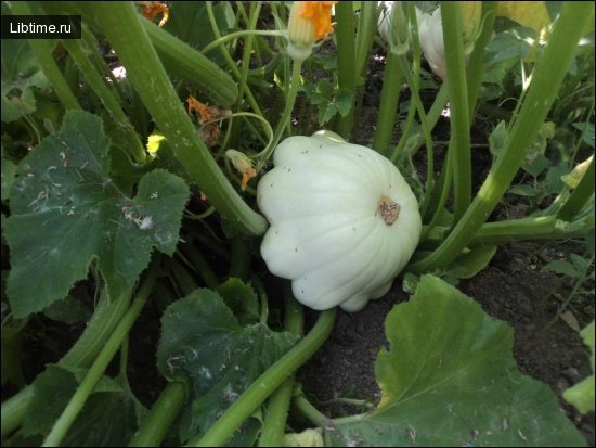
Chromoplasts occur in higher plants: in the petals of flowers, anthers and pistils, fruits, stems, underground organs, sepals, and even in leaves. The appearance and development of chromoplasts is thought to be an adaptive response in plants for attracting insects, birds, and other animals, ensuring pollination and fertilization as well as the dispersal of seeds and fruits in higher archegoniate and flowering plants.
The presence of yellow or orange-red plastids in the underground organs of some cultivated plants is explained by researchers as the result of natural selection acting under the influence of an anthropological factor. 
On the whole, chromoplasts are concentrated in tissues that are ontogenetically relatively older.
What forms do chromoplasts take in plant cells?
Chromoplasts in the cells of higher plants appear not only in the rounded shapes typical of chloroplasts but also as plastids of varied outline. This is especially true of organelles with crystallized pigment. In the cells of red-fleshed watermelon varieties alone, B. T. Matienko was able to count about 42 forms, with needle-shaped and tabular structures predominating.
That author regards some of the minor forms as different stages in the individual differentiation of plastids. There is, however, a view that tabular structures depend on the physico-chemical state of the crystals of the plastids' pigment system. A major role in the formation of these shapes belongs to the physiological environment, which differs across the various parts of the cell.
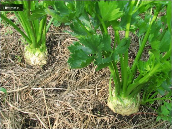
In carrot taproots, E. M. Nedukha counted 32 different forms of crystal-like chromoplasts. W. Straus divided all the coloured plastids of carrot taproots into five basic groups according to shape: filamentous, rounded and curved, polygonal, spiral, and mixed. Filamentous plastids are often found in the cells of root xylem.
Their sizes vary, reaching up to 50–70 µm in length and stretching across the whole cell. Some filaments are straight, while others are twisted in various ways — for example, in a figure-eight shape. Filamentous forms are usually 0.7 µm wide, though even thinner filaments are sometimes encountered at the limit of resolution of the light microscope. The colour intensity of most of the reddish filaments is the same as in the crystal-like bodies.
The group of polygonal chromoplasts is the most numerous in carrot taproots. Among them, quadrangular plates with a rectangular outline are the most common. Viewed from the side, such plastids may look like rhomboids.
Triangular, pentagonal, and hexagonal plates are found less often, as are quadrangular structures with differing angles of inclination. Plastids shaped like spiral ribbons and filaments are also numerous among carrot chromoplasts. This form is the most interesting, since it has no counterpart among objects of inanimate nature.
How does chromoplast shape vary between plant species?
In general, there is a certain specificity in the distribution of particular chromoplast forms across different plant species. Some are dominated by rounded shapes, others by spiral, polygonal, or needle-like forms, and so on.
It should be noted, however, that crystal-like plastids occur more often in the tissues of storage organs, whereas in flower petals chromoplasts of rounded shape are predominantly found.
What is the morphology of amoeboid chromoplasts?
Studying the chromoplasts in the epidermis of the perianth segments of Lilium tigrinum and Tagetes patula, and in the berries of lily of the valley, K. Steffen identified chromoplasts of the amoeboid type. Among them he described primary amoeboid forms, which developed from colourless proplastids or from greened intermediate plastid forms, as well as secondary amoeboid chromoplasts arising from somatic forms after their return to the embryonic state.
Amoeboid chromoplasts have been found in other plants as well. Within a single organ, different plastid forms are observed depending on the histological zonation. In carrot, across separate layers of the secondary cortex of the taproot, E. M. Nedukha observed a clear difference in the shape and size of the plastids and in the state of their pigment system.
In the inner part, adjoining the cambium and consisting of roughly 150 rows of cells, amyloplasts and chromoplasts with starch inclusions (amylochromoplasts) were found. Plastids of rounded shape occur in this zone. Chromoplasts with crystallized pigment were absent here.
In the middle zone, made up of roughly 70 rows of cells, the plastids are represented by amyloplasts, amylochromoplasts, and crystal-like chromoplasts. In the outer zone, consisting of roughly 30 rows of the most differentiated cells, the same types of plastids were also found. Here the number of crystalline chromoplasts and the variability of their forms were greatest.
Apparently, crystal-shaped chromoplasts are more advanced ontogenetically than the rounded amylochromoplasts. Chromoplasts are distributed diffusely throughout the cytoplasm or are concentrated in the central zone around the cell nucleus.
In the regions of the cell adjoining the wall, elongated crystalline plastids are usually oriented parallel to the wall, whereas near the nucleus they point radially toward it. It should be noted that chromoplast studies carried out with light microscopy, despite their abundance, could not form a clear picture of chromoplasts.
Only with the application of electron microscopy methods and biochemical experiments did a deep and comprehensive investigation of chromoplasts begin, since this made it possible to penetrate the fine structure of the organelles and clarify certain aspects of their functioning.